
How to Read an OCT Scan: Understanding Your Eye Test Results
If you have been given an OCT report, you may be wondering how to read an OCT scan when the page is full of unfamiliar pictures, numbers and colour-coded maps. What does an OCT scan show? Does a red or yellow area mean something is wrong? Can I understand the result myself?
An OCT scan can provide remarkably detailed information about the retina, macula and optic nerve. However, an OCT report is not designed to be interpreted as a stand-alone diagnosis. Its meaning depends on the quality of the scan, the eye that was tested, your symptoms, the clinical examination, other tests and how the findings compare with earlier scans.
This article explains how to orient yourself when looking at an OCT eye scan. It does not replace an examination or a doctor’s interpretation.
Important: Seek urgent local medical care for sudden loss or major deterioration of vision, a curtain or shadow in your vision, a sudden increase in flashes or floaters, severe eye pain, a chemical splash or serious injury, or sudden double vision with neurological symptoms. Do not wait for a scheduled online consultation in these situations.
What is an OCT eye test?
If you are wondering what an OCT eye test is, it is a non-invasive imaging test that uses light to produce detailed cross-sectional images of tissues inside the eye, particularly the retina and optic nerve. OCT is sometimes compared with ultrasound because both produce images in slices, although OCT uses light rather than sound.
During an OCT eye test, you usually place your chin and forehead against supports and look at a fixation light while the machine captures images. Nothing normally touches the eye. The scan is usually quick and comfortable, although the exact procedure varies between clinics and some examinations may include dilating drops.
An OCT may be performed as part of an eye examination, to investigate a symptom, or to monitor a known condition over time. It can help an ophthalmologist assess the structure of the eye, but it is only one part of an overall clinical assessment.
What does an OCT eye scan show?
An OCT eye test can provide detailed images of the retina, macula and optic nerve. Depending on the type of scan, an OCT report may show several different views and measurements.
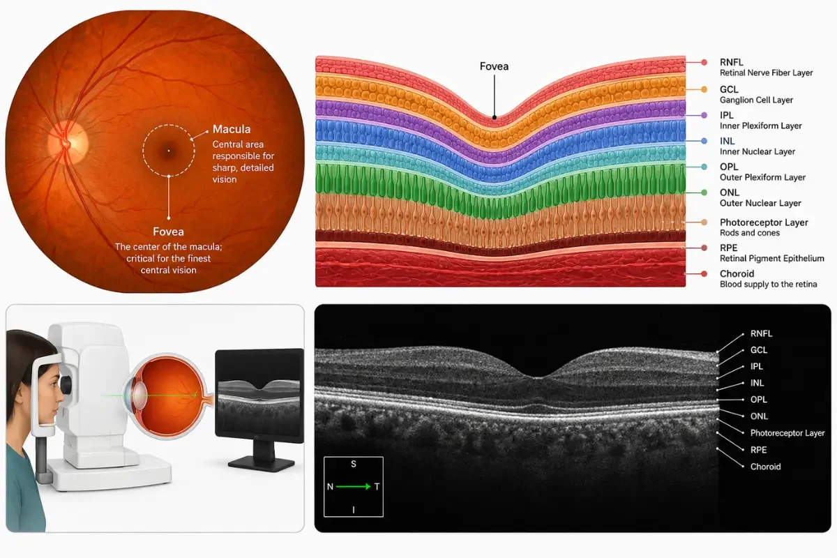
The retinal layers
The retina is a light-sensitive layer at the back of the eye. An OCT cross-section can show the retina as a series of thin layers. The appearance, thickness and organisation of those layers can provide clues about retinal health.
Patients do not need to memorise every retinal layer. The useful question is whether the doctor sees a change in the structure, such as swelling, thinning, a break, a membrane or another abnormal contour.
The macula
The macula is the central part of the retina responsible for detailed central vision. A macular OCT may help assess changes associated with conditions such as age-related macular degeneration, diabetic macular oedema, epiretinal membrane, vitreomacular traction or a macular hole.
An OCT can show features such as thickening, thinning, fluid, tissue distortion or loss of normal structure. These findings still need to be interpreted alongside symptoms and other examination results.
The optic nerve and RNFL
Some OCT scans focus on the optic nerve and the retinal nerve fibre layer, often abbreviated as RNFL. This layer contains nerve fibres that carry visual information towards the brain.
RNFL and related measurements can help assess glaucoma risk and other optic-nerve problems. They do not, by themselves, prove that a person has glaucoma. Eye pressure, the appearance of the optic nerve, visual-field testing, medical history and change over time may all be important.
Thickness maps and measurements
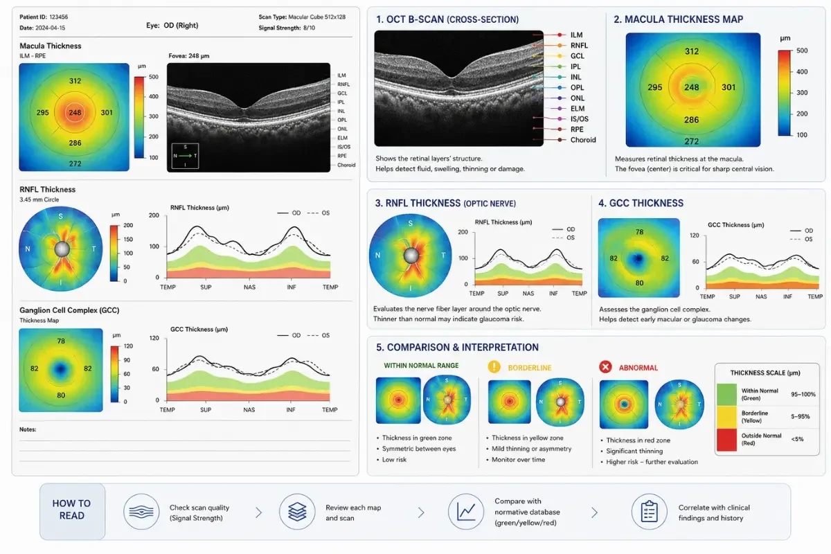
Many reports include measurements in microns, thickness maps, sector charts or trend graphs. These can help the clinician identify areas that are thinner or thicker than expected and monitor change over time.
The number is not meaningful in isolation. Measurements can vary according to the scan protocol, the machine, the position of the scan, the patient’s anatomy and the quality of the image. A result from one device should not automatically be compared with a result from another device.
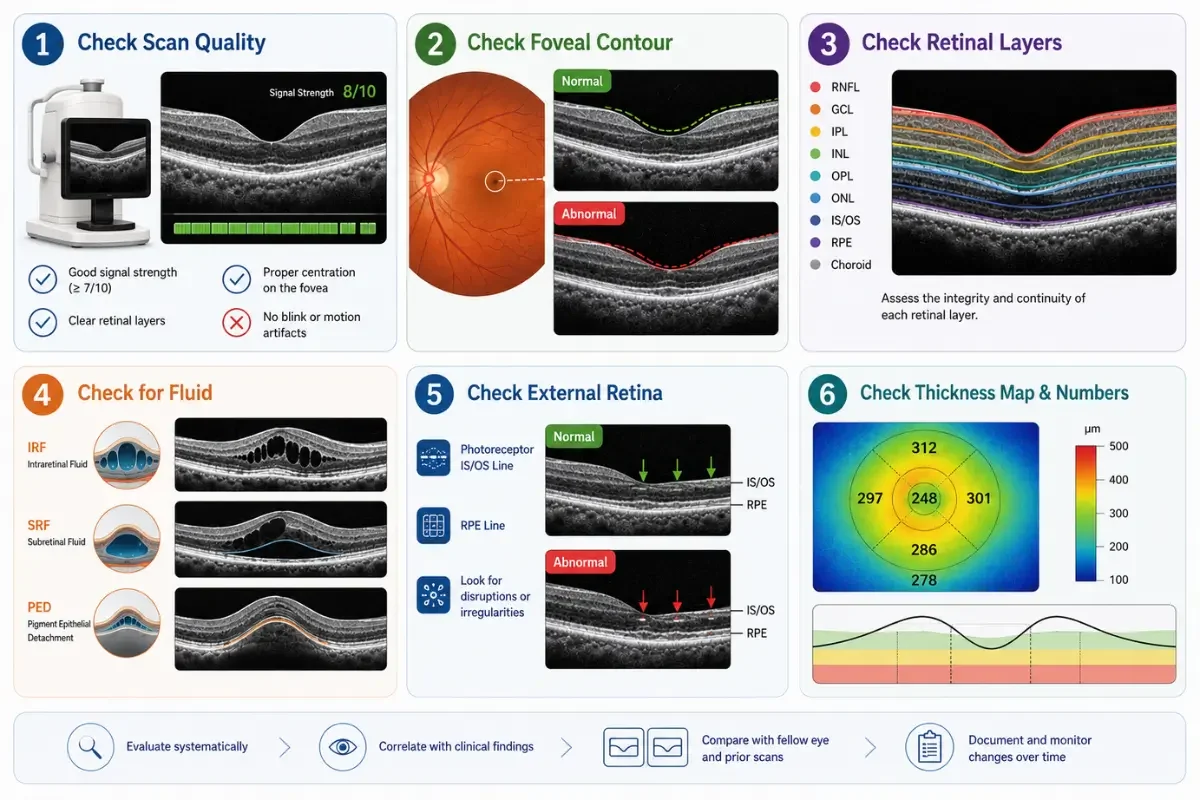
How to read an OCT scan: a patient-level checklist
The following checklist can help you understand the layout of an OCT report and prepare better questions. It is not a method for diagnosing yourself.
1. Check the patient details, date and eye
First confirm that the report belongs to you and that you are looking at the correct eye and examination date. Reports may identify the right and left eye using abbreviations such as OD and OS.
Check whether the report shows a macular scan, optic-nerve scan, RNFL analysis, ganglion-cell analysis or another protocol. Different scan types answer different clinical questions.
If you are comparing reports, make sure you are comparing the same eye and, as far as possible, the same scan area and protocol.
2. Check the scan quality
Scan quality is one of the first things a clinician considers. A report may show a signal-strength score or another quality indicator. The meaning of that score depends on the equipment used.
Poor quality can result from blinking, movement, poor fixation, dry eye, cataract, small pupils or other factors. The machine may also draw segmentation lines incorrectly. These are the lines the software uses to identify the boundaries of retinal layers.
A poor-quality scan can create apparent thinning, thickening or colour changes that do not represent a true disease change. If the image is unclear or the segmentation lines do not follow the tissue correctly, the scan may need to be repeated or reviewed manually.
3. Look at the cross-sectional image, not only the colours
The cross-sectional image is sometimes called a B-scan. It shows a slice through the retina or another area being examined.
The ophthalmologist may look for whether the layers are continuous and appropriately arranged, whether there is fluid or swelling, and whether the normal contour has been changed. They may also look for traction, a membrane, a hole, a scar or other structural features.
Do not assume that every dark or bright area is abnormal. The appearance depends on the tissue, the scan settings and the position of the image. A doctor needs to interpret the pattern in context.
4. Understand what thickness numbers represent
Thickness values usually describe a particular location or region, not the health of the entire eye. A map may divide the macula into rings or sectors, while an optic-nerve report may show a global average and separate quadrants.
The expected range is often based on a reference database. It is not a universal normal value for every person. Age, eye size, refractive error, high myopia, anatomy and other factors can affect the measurement.
For that reason, a small difference from a reference range does not automatically mean that treatment is needed. The pattern and the clinical context matter more than one isolated number.
5. Review RNFL, GCC and optic-nerve information together
An optic-nerve OCT may include RNFL thickness, optic-nerve measurements and sometimes ganglion-cell or ganglion-cell complex analysis, often abbreviated as GCC.
These measurements can support the assessment of glaucoma or another optic-nerve problem. The ophthalmologist may compare the two eyes, examine particular sectors and check whether the result corresponds with the visual-field test.
An apparently low RNFL or GCC value may have several explanations, including natural anatomical variation, high myopia, an unusual optic-nerve shape, scan artefact or true tissue loss. A normal average value also cannot exclude every problem.
6. Treat colour maps as a prompt for review, not a diagnosis
Many OCT reports use colours to compare a measurement with a reference population. Green may indicate that a value is within the device’s expected range, while yellow or red may indicate that it falls closer to or outside that range.
The colours are not a universal traffic-light system. The colour scale can vary between manufacturers, scan types and report pages. A red area does not automatically mean that you have an eye disease, and a green area does not guarantee that every part of the eye is healthy.
Colour results can be affected by scan quality, segmentation errors, high myopia, optic-nerve anatomy and differences between the patient and the reference database. Ask the ophthalmologist what the colour means on your specific report and whether the underlying image supports the software classification.
7. Compare both eyes and previous scans
The two eyes are not always identical, so some asymmetry can be normal. However, a marked difference may be relevant, depending on the pattern and the rest of the examination.
Previous scans are often particularly valuable. A clinician may ask whether a change is real, whether the scans were taken using the same protocol, and whether the change is consistent across several tests. A single result can raise a question; a reliable trend over time may provide stronger evidence.
What do OCT eye test results mean?
OCT reports may describe findings as normal, borderline, outside the expected range, stable or changed. These terms should be read carefully.
Within the expected range: The measurement falls within the reference range used by that device. This does not rule out every eye problem.
Borderline: The result is close to the edge of the reference range. It may need clinical review, repeat testing or comparison with other findings.
Outside the expected range: The measurement differs from the reference database. This is a reason for interpretation, not an automatic diagnosis.
Stable: The clinician has not identified a meaningful change compared with previous reliable tests. Stability is assessed over time and in context.
Changed or progressed: A difference may be important, but it needs confirmation that the scans are comparable and of adequate quality.
Common OCT findings include fluid, swelling, thinning, thickening, traction, a membrane, a hole or a change in the optic-nerve measurements. The same word can have different significance depending on its location and the patient’s history.
If you are unsure what your OCT results mean or whether they need further review, learn more about when to see an eye doctor online and when an online consultation may be appropriate.
What can an OCT scan help detect or monitor?
An OCT scan can help an ophthalmologist evaluate or monitor several types of eye condition, including:
Glaucoma and glaucoma risk: RNFL, GCC and optic-nerve measurements may contribute to the assessment, alongside eye pressure, visual fields and examination findings.
Macular conditions: OCT may show fluid, swelling, tissue distortion, atrophy or other changes associated with age-related macular degeneration and other macular diseases.
Diabetic eye disease: A macular OCT may help identify or monitor retinal swelling associated with diabetes. It does not replace diabetic eye screening or a complete retinal assessment.
Epiretinal membrane and vitreomacular traction: OCT can show distortion or traction affecting the macula.
Macular hole: OCT can help show the structure and extent of a macular hole.
Other retinal or structural abnormalities: The scan may reveal a finding that needs further examination or another type of imaging.
OCT is not a complete eye examination. It does not replace assessment of the front of the eye, eye pressure, visual fields, pupil responses, a dilated examination or other tests when those are needed.
Can you interpret your OCT scan by yourself?
You can learn what the main sections of the report are called, but you should not diagnose yourself from the scan. There are several reasons.
First, the software may produce a misleading result if the scan is poorly centred, blurred or incorrectly segmented. Second, the reference database may not represent your anatomy well. Third, the same appearance can have different meanings in different conditions. Finally, an OCT image does not include all the information obtained from a history and examination.
If a report contains a red or yellow area, the best next step is to ask:
What part of the eye does this section represent?
Is the scan quality good enough to rely on?
Does the underlying image support the colour classification?
Is the finding present in both eyes?
Has it changed compared with earlier scans?
Does it match my symptoms, visual-field test or examination?
Do I need another test or an in-person appointment?
Can an ophthalmologist review an OCT scan online?
Often, yes. An online consultation may be useful when you already have an OCT scan, the written report and a specific question about your diagnosis, monitoring or treatment plan.
During a remote review, an ophthalmologist may be able to:
Explain the terminology and main findings in your report.
Review the underlying OCT images if they are available in a suitable format.
Compare scans from different dates when the images and protocols are adequate.
Discuss how the OCT relates to a known diagnosis or proposed treatment.
Identify information that is missing or needs clarification.
Help you prepare questions for your local eye-care team.
Advise whether a local examination or additional test should be prioritised.
An online consultation cannot perform a new OCT scan, measure eye pressure, carry out a slit-lamp examination, dilate the pupils, perform a visual-field test or provide urgent treatment. It also cannot replace the local ophthalmologist who examines and treats you.
If you want an online OCT review, send the original images and reports where possible rather than only a photograph of a screen. Include the date, the eye tested and any relevant history. Do not send sensitive health information through ordinary social-media messaging; use the secure method provided by the consultation service.
If you already have an OCT scan or report and would like to discuss it with an ophthalmologist, you can book an online eye consultation with Eye Consult Online.
When should you seek urgent local eye care?
Do not wait for an online appointment if you have:
Sudden loss or major deterioration of vision.
A curtain, veil or dark shadow across part of your vision.
A sudden increase in floaters, especially with flashes of light.
Severe eye pain, particularly with redness, headache, nausea or reduced vision.
A chemical splash, penetrating injury or high-speed impact.
A painful red eye while wearing contact lenses.
Sudden double vision together with weakness, facial drooping, speech difficulty, severe headache or other neurological symptoms.
Marked symptoms soon after eye surgery or an eye injection.
Contact a local ophthalmologist, eye emergency service or emergency department promptly. The correct timing depends on the symptom, but sudden or severe changes should be assessed locally rather than managed through a scheduled website consultation.
Frequently asked questions about OCT scans
Is an OCT eye test painful?
OCT is usually non-contact and comfortable. You rest your head and look at a fixation light while the scan is taken. Some appointments may also involve dilating drops or other tests, so ask the clinic what to expect.
How long does an OCT scan take?
The scan itself is usually quick, although the total appointment depends on whether you also need vision testing, photographs, dilation, eye-pressure measurement or a consultation.
Do OCT scans always require dilating drops?
Not always. Whether dilation is needed depends on the machine, the purpose of the examination and the quality of the images required. The clinic should tell you in advance if dilation is planned.
What do the colours on an OCT report mean?
They usually compare a measurement with a reference database used by that machine. The colours are device-specific and should not be interpreted as a diagnosis. The underlying image, scan quality and clinical examination remain important.
Does a red area mean I have an eye disease?
No. A red classification means that a measurement falls outside or near the edge of the device’s reference range. It may reflect disease, normal anatomical variation, high myopia, an artefact or a segmentation error. Ask a clinician to interpret it.
Can an OCT scan diagnose glaucoma?
OCT can provide useful information about the optic nerve, RNFL and GCC, but glaucoma assessment usually combines OCT with eye pressure, visual-field testing, optic-nerve examination, medical history and change over time.
Can an ophthalmologist review an OCT scan from another clinic?
Often, yes. The review is more useful when the original images, written report, dates, eye identification and relevant examination history are available. The ophthalmologist may still recommend local testing if important information is missing.
An OCT scan is evidence, not a verdict
An OCT scan can make the structures inside the eye easier to see, but it does not answer every clinical question by itself. Learning how to read an OCT scan can help you understand the information in your report, but the scan still needs to be interpreted in the context of your symptoms, examination findings, other tests and previous results.
If you are confused by an OCT report, you do not need to become an ophthalmic imaging specialist. You need a clear explanation of what is known, what is uncertain, whether anything has changed and what should happen next.
If you already have an OCT scan or eye-test report and would like professional guidance, learn more about online eye doctor consultations and how an ophthalmologist can review your eye-health information remotely.
Medical disclaimer
This article provides general information and does not replace an individual medical assessment. An OCT scan should be interpreted by a qualified eye-care professional in the context of the complete clinical picture. An online consultation may not be appropriate for every person or every eye problem. Seek urgent local medical care for sudden or severe symptoms.
